How a Revolutionary Microscope Could Reveal Life’s Hidden Cellular Machinery
A new phase for microscopy
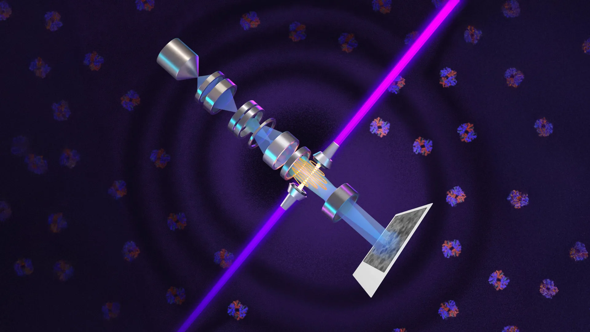
Nearly a century after phase contrast transformed light microscopy, researchers have applied the principle to electron microscopy. The Berkeley physicists’ laser phase plate changes the phase of the microscope’s electron beam, increasing contrast rather than simply lighting the sample.
Electron microscopes can magnify objects roughly 10,000 times more than conventional light microscopes. The new device is designed to improve cryoelectron microscopy, which has advanced protein-structure research and drug discovery but often cannot clearly image very small human proteins.
Seeing smaller proteins
The laser phase plate could allow scientists to image proteins as small as one-third the size of those that challenge current microscopes. It also could make molecules such as hemoglobin easier to distinguish from their surroundings, and improve visibility of cell structures including the nucleus and mitochondria. Holger Müller, the UC Berkeley physics professor who led the development, said the technology is expected to address signal-to-noise limits that prevent many human and animal proteins from being analyzed.
A view inside cells
Cryoelectron tomography combines images taken from multiple angles to build three-dimensional views of proteins and other molecules. Unlike methods that examine isolated molecules in solution, it can show how they are arranged and interact in their natural cellular surroundings.
Bridget Carragher, Biohub’s founding technical director of imaging, said the crowded interior of a cell makes it difficult to locate individual structures. She described the challenge as finding one leaf in a forest and said cryo-ET needs a major improvement in contrast.
Two instruments in development
Carragher is directing construction of a related microscope at Biohub’s Redwood City imaging facility. Her team’s design uses two lasers at right angles, each operating at about half the power, to help prevent components from burning out and reduce optical distortions. Both teams are working with Thermo Fisher Scientific.
Müller called Theia the “Formula 1 microscope,” saying its additional electron optics provide better resolution than standard cryo-EM even without the laser. The Berkeley team’s latest images and technical findings appear in Science.
Key points
- The laser phase plate alters an electron beam to increase image contrast.
- The technology could help image proteins too small for current methods.
- Cryoelectron tomography can show molecules in their natural cellular surroundings.
- Berkeley and Biohub teams are developing microscopes with one- and two-laser designs.